Jacob Light, MD
Abtin Shahlaee, MD
Meera Sivalingam, MD
Wills Eye Hospital
The 2022 ASRS meeting kicked off on Wednesday with the popular case presentations session. There were 24 fascinating medical and surgical cases, which we have divided into Part I and Part II (you can read Part II here). Please enjoy this summary of the cases and key learning points from each.
Case 1: Dr. Vishal Agrawal from SMS hospital presented a patient with persistent vitritis who underwent pars plana vitrectomy. He was found to have a subretinal cyst with contractile elements which was freed from the subretinal space using the cutter. This revealed a cyst with a scolex, consistent with cysticercosis. The cyst was disassembled and removed in the vitreous cavity using the cutter. Despite traditional teaching that disruption of the cyst intraocularly can lead to a robust inflammatory response, Dr. Agrawal highlighted that this is a safe method for complete removal of these cysts which can grow to be quite large, thus making them difficult to remove without disassembly.

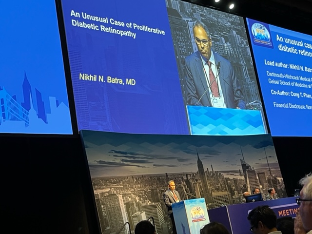
Case 2: Dr. Nikhil Batra from Dartmouth Hitchcock Medical Center then presented a patient with a history of poorly controlled type 1 diabetes who was referred for proliferative diabetic retinopathy. She was found to have severe proliferative diabetic retinopathy. Intravenous fluorescein angiography showed broad posterior pole leakage and foveal avascular zone enlargement. Despite panretinal photocoagulation and intravitreal anti-VEGF, the patient’s disease progressed significantly with widespread macular ischemia. The patient subsequently developed wrist, knee, and ankle pain with associated synovitis. Physical exam showed nail pitting and ridges. Rheumatology diagnosed the patient with psoriatic arthritis and the patient was initiated on Infliximab. Dr. Batra highlighted the importance of a multidisciplinary systemic work up in the setting of an occlusive vasculopathy that fails to improve with traditional proliferative diabetic retinopathy treatments.

Case 3: Dr. Kevin Blinder from The Retina Institute presented a case of an elderly man with recurrent, appositional serous choroidal detachment in the right eye after a glaucoma tube shunt. Drainage was performed using a 27 gauge cannula with anterior placement, just behind iris, allowing for drainage of serous fluid. Direct visualization showed excellent improvement in the choroidal detachment. Dr. Blinder highlighted the value of this minimally invasive method of choroidal drainage.

Case 4: Dr. Avni Finn from Vanderbilt University Medical Center presented patient with a history of S. Caprae associated endophthalmitis one week after cataract surgery who developed multifocal, unilateral white retinal lesions, found to be preretinal on OCT. The lesions improved with observation. Dr. Finn proposed that these lesions were possibly aggregation of the intravitreal vancomycin and ceftazidime versus delayed onset epiretinal inflammation which has been reported in cases of C. acnes endophthalmitis.

Case 5: Dr. Ronald Gentile from New York Eye and Ear Infirmary then presented a case of a middle aged man who was referred for subretinal silicone oil following two prior pars plana vitrectomies. Examination revealed a large optic nerve coloboma. Amniotic membrane was placed over the optic nerve, a retinectomy was created, and PFO was used to remove the subretinal silicone oil. Post operatively, the patient developed macular subretinal fluid, hypothesized to be due to migration of cerebrospinal fluid versus direct communication with the vitreous cavity. On repeat PPV, an amniotic membrane was placed inside the coloboma and the patient had successful resolution of subretinal fluid. Dr. Gentile highlighted the use of amniotic membrane as a barrier against migration of intraocular tamponades and redetachment for optic nerve colobomas

Case 6: Dr. Shrinivas Joshi from MM Joshi Eye Institute presented a young woman who underwent scleral buckle placement for a total retinal detachment. During scleral cut down for external drainage, an altered red reflex was noted and the patient was found to have submacular hemorrhage. The case was converted to a pars plana vitrectomy. The vitreous cutter was introduced via a retinotomy to directly aspirate the submacular hemorrhage. The clot was removed in its entirety and the patient had a successful visual outcome.

Case 7: Dr. M. Ali Khan from Mid Atlantic Retina presented a young boy who presented with left eye pain and redness after working in a woodshop. MRI showed significant orbital inflammation. Examination revealed a linear foreign body embedded in the retina, extending into the vitreous cavity. Intraoperative exploration revealed no scleral laceration or foreign body. A pars plana vitrectomy was performed and the foreign body was removed. Pathology revealed an eyelash! The suspicion is that the patient underwent a high velocity, penetrating injury, carrying the eyelash intraocularly. Take home: never rule out an eyelash in the eye!

Case 8: Dr. Brian Lee then presented a man referred for retinal hemorrhages. Fluorescein angiography revealed neovascularization of the disc and peripheral non-perfusion. Inflammatory systemic workup was negative. The patient underwent panretinal photocoagulation in both eyes. Months later, the patient and his brother were diagnosed with familial amyloidosis. Take home point: amyloidosis can cause proliferative retinopathy, likely due to amyloid deposition within the retinal vasculature. Comments from the audience highlighted that sporadic amyloidosis can be difficult to diagnose, however it is the most common type of systemic amyloidosis.

Case 9: Dr. Jong Park from Associated Retinal Consultants presented middle aged man who underwent a tractional retinal detachment repair. Post operative course was complicated by persistent submacular hemorrhage, elevated IOP and hyphema. Repeat vitrectomy was performed. Under fluid infusion, the surgeon noted indentation and displacement of the subretinal hemorrhage from the infusion jet. The surgeon used the infusion stream to redirect the subretinal hemorrhage away from the macula and drained via a retinal break. Take away: the infusion stream can be used to displace submacular hemorrhage towards an accessible drainage site, allowing for non-traumatic removal of subretinal hemorrhage.

Case 10: Dr. Prethy Rao from Retina and Vitreous of Texas then presented the case of a teenage boy with history of pre B-cell ALL, fusarium sinusitis, oral VZV, and central line infections who presented with bilateral blurred vision for 1-2 weeks. Bilateral peripapillary retinal whitening with extensive hemorrhages were seen on exam. Extensive infectious/oncologic work-up (including MRI and LP) and empiric antimicrobial therapy were negative and ineffective, respectively. The patient’s clinical picture worsened and he developed vitreous hemorrhage without any evidence of vitreous white blood cells. The need for a definitive diagnosis was deemed paramount to guide heme-onc in their decision to proceed with BMT vs palliative care. Diagnostic vitrectomy revealed immature B cells in the vitreous and ocular exam improved with orbital radiation treatment. Take home point: negative MRI and LP does not definitively rule-out presence of intra-ocular lymphoid neoplasia.

Case 11: Dr. Ahmad Rehmani from University of Texas Medical Branch presented a patient who was referred for blurred vision with headache, dizziness, and polyarthralgias. Retinal examination showed bilateral disc edema and multifocal preretinal white lesions. Fluorescein angiography showed a branch arterial occlusion in the left eye and periphlebitis. Careful history revealed the patient recently adopted a stray cat. Bartonella serology was positive and the patient was started on azithromycin with full resolution of disc edema and retinal lesions.

Case 12: Dr. Lincoln Shaw from University of Chicago reported on two female patients who presented with bilateral peripheral retinal hemorrhages and retinal non-perfusion seen on widefield fluorescein angiography. Serologic work-up was consistent with anti-phospholipid syndrome in both cases and systemic anti-coagulation was initiated. He emphasized the utility of widefield imaging in such scenarios.
